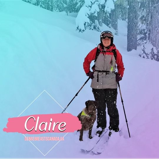
Claire

When the radiologist came into the room carrying a biopsy tray, I was hit by a sense of dread as I was not expecting to see it or him. While preparing a biopsy needle, the radiologist was caring and compassionate. As he disinfected my skin and placed sterile drapes on the area, he asked “Have you ever had an injury to your breast that would create scar tissue in that area?” “Nothing I can recall,” I replied. He then proceeded with the biopsy and, during the time it took to remove four cores of tissue from my breast, he explained that these types of spiculated masses tend to be cancer 90% of the time, and scar tissue in less than 10% of cases. I was shocked, but grateful for the direct and clear statement.
What led to that biopsy? Well, I had a screening mammogram nine months before I found my own lump. My family doctor and I both received my screening mammogram results which were reported as normal. There were no other descriptive words on the template-style report, just “normal”. I was scheduled for mammograms every two years at that point, following recommendations for my age (55).
That “normal” report was the reason that neither of us were particularly concerned or worried when my doctor examined the lump I found but she subsequently ordered a diagnostic mammogram. About a month after I saw my family doctor, I was in the Medical Imaging Department of a local hospital. The mammogram was completed, and then I was escorted by the technologist to the ultrasound room, where another technologist performed that exam. I still was not really worried because, several years earlier, I had needed an ultrasound to confirm a cyst in the same area as this new lump.
It wasn’t until the radiologist came into the room carrying that sterile, green-draped biopsy tray that I knew that my life would be split in two. It would lead to a sense of “before” and an “after”, based on that very day. I had an “interval cancer” which meant that I was diagnosed after my last screening mammogram, before the next one was due. How did this happen?
As a Medical Lab Technologist, I have always found the CBC radio show “White Coat, Black Art” fascinating. I had the feeling that an explanation for my interval cancer might be found in a past episode of that program. I downloaded the podcast and listened to that episode on Dense Breasts, and then I sought my screening mammogram “worksheets” to find out my breast density. I had to make a complaint to the information/privacy commissioner of BC to obtain the documents; it wasn’t easy.
I had regular mammogram screening since I was in my early 40’s, thinking it would lead to early detection, should I ever be one of the 1 in 8 women who experiences this disease. But the most important information from all those years of mammograms was concealed from me and my family doctor. I had category “D” density (the densest) all along. This density increased my risk factor, and if that wasn’t bad enough, the density obscures the images, so the tumour is like “a snowball in a snowstorm”. Supplementary screening, such as ultrasound, can clear away the snowstorm. Breast self-exam is another tool to detect breast cancer in dense breasts.
Several weeks after that last screening mammogram in 2018, the government of BC decided to start informing women and their doctors of mammographic breast density. Access to supplementary screening remains a problem, but at least the era of concealment is over.
In making a surgical decision, I had to take into account that my confidence in reporting and access related to breast cancer screening is pretty much shattered. Could I trust the system to detect cancer in my healthy breast if that occurred? I decided to have a double mastectomy in October 2019. In 2021, after completing the first of many years of anti-hormone therapy, my breast cancer status is “no evidence of active disease”.
