
Cathy
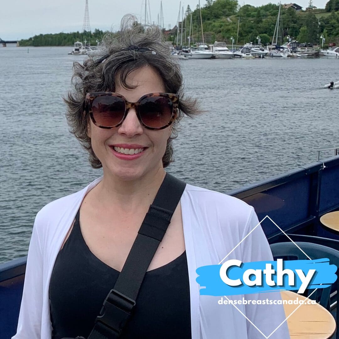
I started going for yearly screening mammograms 12 years ago, at age 38, because my mother had been diagnosed with breast cancer at age 48. Subsequently, my sister was diagnosed with breast cancer at age 46. Both are doing well, and my mother is now in her late 70s.
I thought that by taking a proactive approach to my health, any breast cancer would be detected early. I also asked my doctor about additional screening, such as a breast MRI through the Ontario Breast Screening Program. To determine my eligibility, I met with a genetic counsellor. Through a review of my family and medical history, it was determined that my lifetime risk of developing breast cancer was 21.5%. Since my risk did not exceed 25% and my sister’s genetic testing did not identify BRCA or any other gene mutations, I was advised to continue with yearly mammograms but was not eligible for a supplementary breast MRI. This was disappointing, but I still felt reassured by having yearly mammograms.
My breast density was not factored into the risk assessment. At no point during the years I had been getting mammograms did anyone tell me that I had dense breast tissue or explain what that could mean for my breast cancer risk.
I was diagnosed with breast cancer on February 12, 2025, at age 48, following an abnormal mammogram and biopsy. Two weeks later, I underwent a single mastectomy. Even my surgeon could not feel a lump when he examined me before surgery. At that point, I was sure the tumour was small and had been caught early.
I questioned the need for a mastectomy, and my surgeon explained that the pleomorphic microcalcifications found in my breast, numbering more than 50, were spread throughout the tissue. The only way to remove all the affected tissue while achieving a healthy margin was to remove the entire breast. I asked about a double mastectomy given my family history, but my surgeon did not feel it was necessary at that point. Although a double mastectomy is recommended in some situations, I am glad I did not rush into more extensive surgery. I also decided not to pursue reconstruction because I simply could not put myself through more invasive procedures. It can sometimes be tricky to find clothing that fits properly, but it is something I have learned to accept.
My diagnosis was invasive ductal carcinoma, hormone receptor-positive, stage 3 and grade 3. A higher grade means the cancer cells are unpredictable and are associated with a higher risk of recurrence. Cancer was found in all three sentinel lymph nodes removed during my first surgery. I was devastated to learn that the cancer had already spread to my lymph nodes.
By the time we received the pathology report, a month had already passed since my surgery. From April 2025 onwards, life became a flurry of activity. Numerous diagnostic scans were needed to determine the stage of my cancer, which meant waiting weeks for appointments to be booked. I work in healthcare and understand how things work, but the anxiety of waiting and trying not to think about what the cancer cells might be doing in the meantime was overwhelming.
Fortunately, the scans ruled out cancer in some suspicious areas in my lungs, liver and uterus. However, the PET scan revealed another lymph node containing cancer. The hospital’s tumour board met to discuss my case and determined that chemotherapy needed to happen before addressing the other affected lymph node.
From May to August 2025, I underwent dose-dense chemotherapy every other week for 16 weeks. The regimen was AC-T: doxorubicin and cyclophosphamide for 4 cycles, followed by paclitaxel for the remaining 4 cycles. After a month of recovery, axillary dissection surgery was scheduled for September 2025 to remove the remaining axillary lymph nodes. Although clearing the nodes increases the risk of lymphedema, my surgeon explained that the surgery would give me the best chance of preventing further spread of the cancer.
In total, nine lymph nodes were removed during the second surgery, and cancer was found in two more nodes. I was told that it’s possible there was an incomplete response to chemotherapy, since the cancer was still present in the nodes after treatment. While it is uncertain why this happens, it can occur with grade 3 cancer due to its unpredictable nature. It doesn’t mean the treatment had zero effect. How much of an effect it had is simply unknown. It was very upsetting to hear this because I had kept reassuring myself that the treatment would mop up any cancer cells circulating in my body.
My oncologist stressed that we need to take an aggressive approach over the next two years to target any remaining cancer cells and reduce the risk of recurrence. I was advised to start hormone therapy (Letrozole) for eight years, monthly ovarian suppression injections (Zoladex), and targeted therapy (Verzenio) for two years. The targeted therapy works differently from hormone therapy, providing another layer of protection against recurrence. I will also receive intravenous bone-strengthening treatment (Zometa) twice per year for several years. This will strengthen my bones and prevent fractures, as some of the medications can impact bone density, while also further reducing the risk of cancer recurrence.
Before my cancer diagnosis, I had never taken medication for anything, so this was a significant mental hurdle for me. While chemotherapy was relatively short-term, it still left behind lingering long-term side effects. These new ongoing medications bring daily side effects of their own, serving as a constant reminder of my cancer journey. I started the medications in October 2025, which were all phased in one by one.
In December 2025, I completed radiation therapy. My treatment was delivered on an accelerated schedule of six intensive sessions, which carried the clinical equivalent dose of fifteen traditional treatments.
When I was first diagnosed, my oncologists were urgently focused on treating the cancer. However, I was still questioning why it had not been caught sooner, especially given how regularly I had undergone screening mammograms. I thought I was doing everything right, but you don’t know what you don’t know.
I had never asked for copies of my mammogram reports because I did not think I needed to. After my diagnosis, I reviewed the reports and noticed some key terms: heterogeneously dense, extremely dense, and Category C. I learned that breast density can change over time, which was reflected in my results. Regardless, my breasts had always fallen into a dense-breast category. Even though my scans showed no evidence of malignancy, dense tissue meant that cancer could still be present but remain unseen on a mammogram. This was never explained to me.
I was angry to realize that knowing my breast density sooner might have prompted me to ask about additional screening, such as ultrasound alongside mammography. Although I was not eligible for breast MRI, an ultrasound might have detected the cancer earlier, potentially resulting in a lower risk of recurrence, fewer treatments and fewer surgeries. Not to mention the potential savings to the healthcare system associated with avoiding a stage 3 diagnosis.
Even now, it is challenging to advocate for ultrasound screening with my doctor. I was grateful to find the Dense Breasts Canada website and all the information that I could use when discussing my concerns with my doctor. It is deeply upsetting to go through something like this and still feel that I have to push and advocate for these tests. However, I do not blame any single doctor. Rather, I see this as a sign that we have a lot of work to do to ensure that healthcare professionals and patients are up to date on the risks associated with dense breasts. People need the information necessary to make informed decisions about screening and to advocate for themselves.
I hope sharing my story helps anyone facing this risk avoid going through the same experience.